Questions and Answers ​in MRI
  • Home
  • Complete List of Questions
  • …Magnets & Scanners
    • Basic Electromagnetism >
      • What causes magnetism?
      • What is a Tesla?
      • Who was Tesla?
      • What is a Gauss?
      • How strong is 3.0T?
      • What is a gradient?
      • Aren't gradients coils?
      • What is susceptibility?
      • How to levitate a frog?
      • What is ferromagnetism?
      • Superparamagnetism?
    • Magnets - Part I >
      • Types of magnets?
      • Brands of scanners?
      • Which way does field point?
      • Which is the north pole?
      • Low v mid v high field?
      • Advantages to low-field?
      • Disadvantages?
      • What is homogeneity?
      • Why homogeneity?
      • Why shimming?
      • Passive shimming?
      • Active shimming?
    • Magnets - Part II >
      • Superconductivity?
      • Perpetual motion?
      • How to ramp?
      • Superconductive design?
      • Room Temp supercon?
      • Liquid helium use?
      • What is a quench?
      • Is field ever turned off?
      • Emergency stop button?
    • Gradients >
      • Gradient coils?
      • How do z-gradients work?
      • X- and Y- gradients?
      • Open scanner gradients?
      • Eddy current problems?
      • Active shielded gradients?
      • Active shield confusion?
      • What is pre-emphasis?
      • Gradient heating?
      • Gradient specifications?
      • Gradient linearity?
    • RF & Coils >
      • Many kinds of coils?
      • Radiofrequency waves?
      • Phase v frequency?
      • RF Coil function(s)?
      • RF-transmit coils?
      • LP vs CP (Quadrature)?
      • Multi-transmit RF?
      • Receive-only coils?
      • Array coils?
      • AIR Coils?
    • Site Planning >
      • MR system layout?
      • What are fringe fields?
      • How to reduce fringe?
      • Magnetic shielding?
      • Need for vibration testing?
      • What's that noise?
      • Why RF Shielding?
      • Wires/tubes thru wall?
  • ...Safety and Screening
    • Overview >
      • ACR Safety Zones?
      • MR safety screening?
      • Incomplete screening?
      • Passive v active implants?
      • Conditional implants?
      • Common safety issues?
      • Projectiles?
      • Metal detectors?
      • Pregnant patients?
      • Postop, ER & ICU patients?
      • Temperature monitoring?
      • Orbital foreign bodies?
      • Bullets and shrapnel?
    • Static Fields >
      • "Dangerous" metals?
      • "Safe" metals?
      • Magnetizing metal?
      • Object shape?
      • Forces on metal?
      • Most dangerous place?
      • Force/torque testing?
      • Static field bioeffects?
      • Dizziness/Vertigo?
      • Flickering lights?
      • Metallic taste?
    • RF Fields >
      • RF safety overview?
      • RF biological effects?
      • What is SAR?
      • SAR limits?
      • Operating modes?
      • How to reduce SAR?
      • RF burns?
      • Estimate implant heating?
      • SED vs SAR?
      • B1+rms vs SAR?
      • Personnel exposure?
      • Cell phones?
    • Gradient Fields >
      • Gradient safety overview
      • Acoustic noise?
      • Nerve stimulation?
      • Gradient vs RF heating?
    • Safety: Neurological >
      • Aneurysm coils/clips?
      • Shunts/drains?
      • Pressure monitors/bolts?
      • Deep brain stimulators?
      • Spinal cord stimulators?
      • Vagal nerve stimulators?
      • Cranial electrodes?
      • Carotid clamps?
      • Peripheral stimulators?
      • Epidural catheters?
    • Safety: Head & Neck >
      • Additional orbit safety?
      • Cochlear Implants?
      • Bone conduction implants?
      • Other ear implants?
      • Dental/facial implants?
      • ET tubes & airways?
    • Safety: Chest & Vascular >
      • Breast tissue expanders?
      • Breast biopsy markers?
      • Airway stents/valves/coils?
      • Respiratory stimulators?
      • Ports/vascular access?
      • Swan-Ganz catheters?
      • IVC filters?
      • Implanted infusion pumps?
      • Insulin pumps & CGMs?
      • Vascular stents/grafts?
      • Sternal wires/implants?
    • Safety: Cardiac >
      • Pacemaker dangers?
      • Pacemaker terminology?
      • New/'Safe" Pacemakers?
      • Old/Legacy Pacemakers?
      • Violating the conditions?
      • Epicardial pacers/leads?
      • Cardiac monitors?
      • Heart valves?
      • Miscellaneous CV devices?
    • Safety: Abdominal >
      • PIllCam and capsules?
      • Gastric pacemakers?
      • Other GI devices?
      • Contraceptive devices?
      • Foley catheters?
      • Incontinence devices?
      • Penile Implants?
      • Sacral nerve stimulators?
      • GU stents and other?
    • Safety: Orthopedic >
      • Orthopedic hardware?
      • External fixators?
      • Traction and halos?
      • Bone stimulators?
      • Magnetic rods?
  • …The NMR Phenomenon
    • Spin >
      • What is spin?
      • Why I = ½, 1, etc?
      • Proton = nucleus = spin?
      • Predict nuclear spin (I)?
      • Magnetic dipole moment?
      • Gyromagnetic ratio (γ)?
      • "Spin" vs "Spin state"?
      • Energy splitting?
      • Fall to lowest state?
      • Quantum "reality"?
    • Precession >
      • Why precession?
      • Who was Larmor?
      • Energy for precession?
      • Chemical shift?
      • Net magnetization (M)?
      • Does M instantly appear?
      • Does M also precess?
      • Does precession = NMR?
    • Resonance >
      • MR vs MRI vs NMR?
      • Who discovered NMR?
      • How does B1 tip M?
      • Why at Larmor frequency?
      • What is flip angle?
      • Spins precess after 180°?
      • Phase coherence?
      • Release of RF energy?
      • Rotating frame?
      • Off-resonance?
      • Adiabatic excitation?
      • Adiabatic pulses?
    • Relaxation - Physics >
      • Bloch equations?
      • What is T1?
      • What is T2?
      • Relaxation rate vs time?
      • Why is T1 > T2?
      • T2 vs T2*?
      • Causes of Relaxation?
      • Dipole-dipole interactions?
      • Chemical Exchange?
      • Spin-Spin interactions?
      • Macromolecule effects?
      • Which H's produce signal?
      • "Invisible" protons?
      • Magnetization Transfer?
      • Bo effect on T1 & T2?
      • How to predict T1 & T2?
    • Relaxation - Clincial >
      • T1 bright? - fat
      • T1 bright? - other oils
      • T1 bright? - cholesterol
      • T1 bright? - calcifications
      • T1 bright? - meconium
      • T1 bright? - melanin
      • T1 bright? - protein/mucin
      • T1 bright? - myelin
      • Magic angle?
      • MT Imaging/Contrast?
  • …Pulse Sequences
    • MR Signals >
      • Origin of MR signal?
      • Free Induction Decay?
      • Gradient echo?
      • TR and TE?
      • Spin echo?
      • 90°-90° Hahn Echo?
      • Stimulated echoes?
      • STEs for imaging?
      • 4 or more RF-pulses?
      • Partial flip angles?
      • How is signal higher?
      • Optimal flip angle?
    • Spin Echo >
      • SE vs Multi-SE vs FSE?
      • Image contrast: TR/TE?
      • Opposite effects ↑T1 ↑T2?
      • Meaning of weighting?
      • Does SE correct for T2?
      • Effect of 180° on Mz?
      • Direction of 180° pulse?
    • Inversion Recovery >
      • What is IR?
      • Why use IR?
      • Phase-sensitive IR?
      • Why not PSIR always?
      • Choice of IR parameters?
      • TI to null a tissue?
      • STIR?
      • T1-FLAIR
      • T2-FLAIR?
      • IR-prepped sequences?
      • Double IR?
    • Gradient Echo >
      • GRE vs SE?
      • Multi-echo GRE?
      • Types of GRE sequences?
      • Commercial Acronyms?
      • Spoiling - what and how?
      • Spoiled-GRE parameters?
      • Spoiled for T1W only?
      • What is SSFP?
      • GRASS/FISP: how?
      • GRASS/FISP: parameters?
      • GRASS vs MPGR?
      • PSIF vs FISP?
      • True FISP/FIESTA?
      • FIESTA v FIESTA-C?
      • DESS?
      • MERGE/MEDIC?
      • GRASE?
      • MP-RAGE v MR2RAGE?
    • Susceptibility Imaging >
      • What is susceptibility (χ)?
      • What's wrong with GRE?
      • Making an SW image?
      • Phase of blood v Ca++?
      • Quantitative susceptibility?
    • Diffusion: Basic >
      • What is diffusion?
      • Iso-/Anisotropic diffusion?
      • "Apparent" diffusion?
      • Making a DW image?
      • What is the b-value?
      • b0 vs b50?
      • Trace vs ADC map?
      • Light/dark reversal?
      • T2 "shine through"?
      • Exponential ADC?
      • T2 "black-out"?
      • DWI bright causes?
    • Diffusion: Advanced >
      • Diffusion Tensor?
      • DTI (tensor imaging)?
      • Whole body DWI?
      • Readout-segmented DWI?
      • Small FOV DWI?
      • IVIM?
      • Diffusion Kurtosis?
    • Fat-Water Imaging >
      • Fat & Water properties?
      • F-W chemical shift?
      • In-phase/out-of-phase?
      • Best method?
      • Dixon method?
      • "Fat-sat" pulses?
      • Water excitation?
      • STIR?
      • SPIR?
      • SPAIR v SPIR?
      • SPIR/SPAIR v STIR?
  • …Making an Image
    • From Signals to Images >
      • Phase v frequency?
      • Angular frequency (ω)?
      • Signal squiggles?
      • Real v Imaginary?
      • Fourier Transform (FT)?
      • What are 2D- & 3D-FTs?
      • Who invented MRI?
      • How to locate signals?
    • Frequency Encoding >
      • Frequency encoding?
      • Receiver bandwidth?
      • Narrow bandwidth?
      • Slice-selective excitation?
      • SS gradient lobes?
      • Cross-talk?
      • Frequency encode all?
      • Mixing of slices?
      • Two slices at once?
      • Simultaneous Multi-Slice?
    • Phase Encoding >
      • Phase-encoding gradient?
      • Single PE step?
      • What is phase-encoding?
      • PE and FE together?
      • 2DFT reconstruction?
      • Choosing PE/FE direction?
    • Performing an MR Scan >
      • What are the steps?
      • Automatic prescan?
      • Routine shimming?
      • Coil tuning/matching?
      • Center frequency?
      • Transmitter gain?
      • Receiver gain?
      • Dummy cycles?
      • Where's my data?
      • MR Tech qualifications?
    • Image Quality Control >
      • Who regulates MRI?
      • Who accredits?
      • Mandatory accreditation?
      • Routine quality control?
      • MR phantoms?
      • Geometric accuracy?
      • Image uniformity?
      • Slice parameters?
      • Image resolution?
      • Signal-to-noise?
      • Ghosting?
  • …K-space & Rapid Imaging
    • K-space (Basic) >
      • What is k-space?
      • Parts of k-space?
      • What does "k" stand for?
      • Spatial frequencies?
      • Locations in k-space?
      • Data for k-space?
      • Why signal ↔ k-space?
      • Spin-warp imaging?
      • Big spot in middle?
      • K-space trajectories?
      • Radial sampling?
    • K-space (Advanced) >
      • K-space grid?
      • Negative frequencies?
      • Field-of-view (FOV)
      • Rectangular FOV?
      • Partial Fourier?
      • Phase symmetry?
      • Read symmetry?
      • Why not use both?
      • ZIP?
    • Rapid Imaging (FSE &EPI) >
      • What is FSE/TSE?
      • FSE parameters?
      • Bright Fat?
      • Other FSE differences?
      • Dual-echo FSE?
      • Driven equilibrium?
      • Reduced flip angle FSE?
      • Hyperechoes?
      • SPACE/CUBE/VISTA?
      • Echo-planar imaging?
      • HASTE/SS-FSE?
    • Parallel Imaging (PI) >
      • What is PI?
      • How is PI different?
      • PI coils and sequences?
      • Why and when to use?
      • Two types of PI?
      • SENSE/ASSET?
      • GRAPPA/ARC?
      • CAIPIRINHA?
      • Compressed sensing?
      • Noise in PI?
      • Artifacts in PI?
  • …Contrast Agents
    • Contrast Agents: Physics >
      • Why Gadolinium?
      • Paramagnetic relaxation?
      • What is relaxivity?
      • Why does Gd shorten T1?
      • Does Gd affect T2?
      • Gd & field strength?
      • Best T1-pulse sequence?
      • Triple dose and MT?
      • Dynamic CE imaging?
      • Gadolinium on CT?
    • Contrast Agents: Clinical >
      • So many Gd agents!
      • Important properties?
      • Ionic v non-ionic?
      • Intra-articular/thecal Gd?
      • Gd liver agents (Eovist)?
      • Mn agents (Teslascan)?
      • Feridex & Liver Agents?
      • Lymph node agents?
      • Ferumoxytol?
      • Blood pool (Ablavar)?
      • Bowel contrast agents?
    • Contrast Agents: Safety >
      • Gadolinium safety?
      • Allergic reactions?
      • Renal toxicity?
      • What is NSF?
      • NSF by agent?
      • Informed consent for Gd?
      • Gd protocol?
      • Is Gd safe in infants?
      • Reduced dose in infants?
      • Gd in breast milk?
      • Gd in pregnancy?
      • Gd accumulation?
      • Gd deposition disease?
  • …Cardiovascular and MRA
    • Flow effects in MRI >
      • Defining flow?
      • Expected velocities?
      • Laminar v turbulent?
      • Predicting MR of flow?
      • Time-of-flight effects?
      • Spin phase effects?
      • Flow void?
      • Why GRE ↑ flow signal?
      • Slow flow v thrombus?
      • Even-echo rephasing?
      • Flow-compensation?
      • Flow misregistration?
    • MR Angiography - I >
      • MRA methods?
      • Dark vs bright blood?
      • Time-of-Flight (TOF) MRA?
      • 2D vs 3D MRA?
      • MRA parameters?
      • Magnetization Transfer?
      • Ramped flip angle?
      • MOTSA?
      • Fat-suppressed MRA?
      • TOF MRA Artifacts?
      • Phase-contrast MRA?
      • What is VENC?
      • Measuring flow?
      • 4D Flow Imaging?
      • How accurate?
    • MR Angiography - II >
      • Gated 3D FSE MRA?
      • 3D FSE MRA parameters?
      • SSFP MRA?
      • Inflow-enhanced SSFP?
      • MRA with ASL?
      • Other MRA methods?
      • Contrast-enhanced MRA?
      • Timing the bolus?
      • View ordering in MRA?
      • Bolus chasing?
      • TRICKS or TWIST?
      • CE-MRA artifacts?
    • Cardiac I - Intro/Anatomy >
      • Cardiac protocols?
      • Patient prep?
      • EKG problems?
      • Magnet changes EKG?
      • Gating v triggering?
      • Gating parameters?
      • Heart navigators?
      • Dark blood/Double IR?
      • Why not single IR?
      • Triple IR?
      • Polar plots?
      • Coronary artery MRA?
    • Cardiac II - Function >
      • Beating heart movies?
      • Cine parameters?
      • Real-time cine?
      • Ventricular function?
      • Tagging/SPAMM?
      • Perfusion: why and how?
      • 1st pass perfusion?
      • Quantifying perfusion?
      • Dark rim artifact
    • Cardiac III - Viability >
      • Gd enhancement?
      • TI to null myocardium?
      • PS (phase-sensitive) IR?
      • Wideband LGE?
      • T1 mapping?
      • Iron/T2*-mapping?
      • Edema/T2-mapping?
      • Why/how stress test?
      • Stess drugs/agents?
      • Stress consent form?
  • …MR Artifacts
    • Tissue-related artifacts >
      • Chemical shift artifact?
      • Chemical shift in phase?
      • Reducing chemical shift?
      • Chemical Shift 2nd Kind?
      • In-phase/out-of phase?
      • IR bounce point?
      • Susceptibility artifact?
      • Metal suppression?
      • Dielectric effect?
      • Dielectric Pads?
    • Motion-related artifacts >
      • Why discrete ghosts?
      • Motion artifact direction?
      • Reducing motion artifacts?
      • Saturation pulses?
      • Gating methods?
      • Respiratory comp?
      • Navigator echoes?
      • PROPELLER/BLADE?
    • Technique-related artifacts >
      • Partial volume effects?
      • Slice overlap?
      • Aliasing?
      • Wrap-around artifact?
      • Eliminate wrap-around?
      • Phase oversampling?
      • Frequency wrap-around?
      • Spiral/radial artifacts?
      • Gibbs artifact?
      • Nyquist (N/2) ghosts?
      • Zipper artifact?
      • Data artifacts?
      • Surface coil flare?
      • MRA Artifacts (TOF)?
      • MRA artifacts (CE)?
  • …Functional Imaging
    • Perfusion I: Intro & DSC >
      • Measuring perfusion?
      • Meaning of CBF, MTT etc?
      • DSC v DCE v ASL?
      • How to perform DSC?
      • Bolus Gd effect?
      • T1 effects on DSC?
      • DSC recirculation?
      • DSC curve analysis?
      • DSC signal v [Gd]
      • Arterial input (AIF)?
      • Quantitative DSC?
    • Perfusion II: DCE >
      • What is DCE?
      • How is DCE performed?
      • How is DCE analyzed?
      • Breast DCE?
      • DCE signal v [Gd]
      • DCE tissue parmeters?
      • Parameters to images?
      • K-trans = permeability?
      • Utility of DCE?
    • Perfusion III: ASL >
      • What is ASL?
      • ASL methods overview?
      • CASL?
      • PASL?
      • pCASL?
      • ASL parameters?
      • ASL artifacts?
      • Gadolinium and ASL?
      • Vascular color maps?
      • Quantifying flow?
    • Functional MRI/BOLD - I >
      • Who invented fMRI?
      • How does fMRI work?
      • BOLD contrast?
      • Why does BOLD ↑ signal?
      • Does BOLD=brain activity?
      • BOLD pulse sequences?
      • fMRI Paradigm design?
      • Why "on-off" comparison?
      • Motor paradigms?
      • Visual?
      • Language?
    • Functional MRI/BOLD - II >
      • Process/analyze fMRI?
      • Best fMRI software?
      • Data pre-processing?
      • Registration/normalization?
      • fMRI statistical analysis?
      • General Linear Model?
      • Activation "blobs"?
      • False activation?
      • Resting state fMRI?
      • Analyze RS-fMRI?
      • Network/Graphs?
      • fMRI at 7T?
      • Mind reading/Lie detector?
      • fMRI critique?
  • …MR Spectroscopy
    • MRS I - Basics >
      • MRI vs MRS?
      • Spectra vs images?
      • Chemical shift (δ)?
      • Measuring δ?
      • Backward δ scale?
      • Predicting δ?
      • Size/shapes of peaks?
      • Splitting of peaks?
      • Localization methods?
      • Single v multi-voxel?
      • PRESS?
      • STEAM?
      • ISIS?
      • CSI?
    • MRS II - Clinical ¹H MRS >
      • How-to: brain MRS?
      • Water suppression?
      • Fat suppression?
      • Normal brain spectra?
      • Choice of TR/TE/etc?
      • Hunter's angle?
      • Lactate inversion?
      • Metabolite mapping?
      • Metabolite quantitation?
      • Breast MRS?
      • Gd effect on MRS?
      • How-to: prostate MRS?
      • Prostate spectra?
      • Muscle ¹H-MRS?
      • Liver ¹H-MRS?
      • MRS artifacts?
    • MRS III - Multi-nuclear >
      • Other nuclei?
      • Why phosphorus?
      • How-to: ³¹P MRS
      • Normal ³¹P spectra?
      • Organ differences?
      • ³¹P measurements?
      • Decoupling?
      • NOE?
      • Carbon MRS?
      • Sodium imaging?
      • Xenon imaging?
  • ...Artificial Intelligence
    • AI Part I: Basics >
      • Artificial Intelligence (AI)?
      • What is a neural network?
      • Machine Learning (ML)?
      • Shallow v Deep ML?
      • Shallow networks?
      • Deep network types?
      • Data prep and fitting?
      • Back-Propagation?
      • DL 'Playground'?
    • AI Part 2: Advanced >
      • What is convolution?
      • Convolutional Network?
      • Softmax?
      • Upsampling?
      • Limitations/Problems of AI?
      • Is the Singularity near?
    • AI Part 3: Image processing >
      • AI in clinical MRI?
      • Super-resolution?
  • ...Tissue Properties Imaging
    • MRI of Hemorrhage >
      • Hematoma overview?
      • Types of Hemoglobin?
      • Hyperacute/Oxy-Hb?
      • Acute/Deoxy-Hb?
      • Subacute/Met-Hb?
      • Deoxy-Hb v Met-Hb?
      • Extracellular met-Hb?
      • Chronic hematomas?
      • Hemichromes?
      • Ferritin/Hemosiderin?
      • Subarachnoid blood?
      • Blood at lower fields?
    • T2 cartilage mapping
    • MR Elastography?
    • Synthetic MRI?
    • Amide Proton Transfer?
    • MR thermography?
    • Electric Properties Imaging?
  • Copyright/Legal
    • Copyright Issues
    • Legal Disclaimers
  • Forums/Blogs/Links
  • What's New
  • Self-test Quizzes - NEW!
    • Magnets & Scanners Quiz
    • Safety & Screening Quiz
    • NMR Phenomenon Quiz
    • Pulse Sequences Quiz
    • Making an Image Quiz
    • K-space & Rapid Quiz
    • Contrast & Blood Quiz
    • Cardiovascular & MRA Quiz

Spin Echo (SE)

How is a spin echo generated?  
Spin Echo (SE)
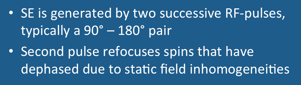
A single RF pulse generates a free induction decay (FID), but two successive RF pulses produce a spin echo (SE).  The time between the middle of the first RF pulse and the peak of the spin echo is called the echo time (TE).  

This remarkable discovery was made in 1949 by Erwin Hahn and can be counted among the most important developments in the history of NMR.  But where does the SE come from?
Picture
Creation of a spin echo. by two RF pulses. The first RF pulse generates an FID, while the second pulse generates the SE. The echo time (TE) is twice the interpulse interval.
Erwin HahnErwin Hahn (1921-2016)
The SE represents regeneration of spin phase information apparently lost during the decay of the FID. The "rebirth" of the FID as a SE is possible because many of the T2* processes which originally produced the decay of the FID are symmetrically reversible.  In fact, most of the FID signal has not been destroyed;  it has merely become "disorganized" because the individual spins comprising it have lost their phase coherence. The system is said to possess a "hidden order" or "atomic memory".  By applying a second RF pulse, certain dephased components of the original FID can be refocused into a SE.  

The animation below shows how a spin echo is formed.  The red arrows represent groups of spins that experience identical local fields and respond similarly. (Such groups of like-behaving spins are called spin isochromats, literally "spins of the same color".)

The 90°-pulse first tips these spins into the transverse plane.  Because the local microscopic fields may differ slightly, some spin groups may precess faster (and gain phase) relative to others. This is represented by the spreading out of arrows in the transverse plane. The faster spins initially rotate toward the viewer and the slower spins rotate away.
Spin Echo (SE)
Formation of the spin echo from Wikipedia, created by
Gavin W. Morley. Click on image for animation.
The 180°-pulse now turns the entire system on its head. An analogy to "flipping a pancake" is sometimes made. After the flip, the faster precessing spins now find themselves at the back of the pack. With continued evolution they eventually catch up with the slower spins. This occurs at time TE = 2 x t  which is the center of the spin echo. Beyond the echo center the faster spins once again leave the slower ones behind and the system again dephases.
Although the example above shows the generation of spin echoes by successive 90° and 180° pulses, these precise flip angles are not required.  In fact, spin echoes are formed when two successive RF-pulses of any flip angle are employed! Hahn, in his original paper, used two 90° pulses. When flip angles other than 90° and 180° are employed, the resultant spin echo is sometimes referred to as a Hahn echo.
 

Advanced Discussion (show/hide)»

No supplementary material yet. Check back soon!

References
     Brewer RG, Hahn EL. Atomic memory. Sci Amer 1984:251(6):50-57.
     Feinberg DA. The transformative genius of Erwin Hahn. Magnetic Resonance in Medicine Highlights, May 2016; pp. 2-9. (an interview of the 95-year-old Dr. Hahn shortly before his death loaded with interesting insights about his discoveries and the early days of NMR) 
     Hahn EL. Spin echoes. Phys Rev 1950;80:580-594.
     "Spin Echo". Wikipedia. The Free Encyclopedia.

Related Questions
    Where does the MR signal come from? 
    What is the difference between spin echo, multi-spin echo and fast spin echo? 


←  Previous Question
Next Question  →
↑ Complete List of Questions ↑
© 2024 AD Elster, ELSTER LLC
All rights reserved.   
MRIquestions.com - Home
Donate
Please help keep this site free for everyone in the world!