Phase-Contrast MRAHow does phase-contrast MR angiography work?
|
|
The idea that blood flow velocities could be encoded by phase was first developed by my mentor, Paul R. ("Dick") Moran in the early 1980's. Dick had trained under the great Charlie Slichter, a famous researcher at University of Illinois at Urbana-Champaign who had discovered J-coupling and was the author of the most widely read textbook in the field for several decades. Charlie, in turn, had been trained by Nobel Prize winner Ed Purcell. So that's my lineage back to NMR royalty!
|
Moran analyzed the phase effects on stationary and moving spins subjected to a pair of bipolar gradients as illustrated in the diagram right. A stationary spin subjected to such a gradient pair will experience no net phase shift, but a moving spin will have a net phase shift proportional to its velocity. Two spins flowing at the same speed but in opposite directions will have equal but opposite phase shifts. By measuring changes in phase, therefore, velocity can be computed.
|
Effect of bipolar phase-encoding gradients.
Net phase gain is zero for stationary spins but positive for spins moving in the direction of the gradients. |
Usually bipolar gradients are applied along only one anatomic axis (x-, y-, or z-) at a time to provide flow sensitivity in that specific direction. However, flow-encoding gradients may be applied along two or more axes simultaneously to measure flow sensitivity along any arbitrary direction. This method will not produce an angiographic image but is the basis for phase contrast flow measurements widely used in cardiovascular MRI and discussed in detail in subsequent Q&A's.
The amplitude, duration, and spacing of the bipolar gradients determine the degree of sensitivity to slow or fast flows. This is controlled by an operator-selectable parameter known as velocity encoding (VENC) which must be prescribed prior to any PC MRI/MRA study. Proper setting of VENC is critical to the performance of the study and is the subject of a separate Q&A.
By applying bipolar gradients sequentially along the cardinal directions (x-, y-, and z-) and measuring phase shifts, velocity components of flow along each direction (vx, vy, and vz) can be computed.
The flow data obtained from a PC study may obtained in 1D, 2D, 3D, or 4D mode. It can be used not only for blood flow measurements, but to depict the flow of any fluid such as CSF. It can be displayed in various ways: as a directionally-insensitive magnitude ("speed") image similar to a TOF MRA; in a Doppler-like directionally sensitive color- or gray-scale format; or even in cine mode.
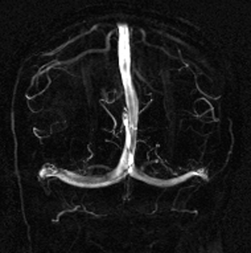
2D PC MR venogram
|
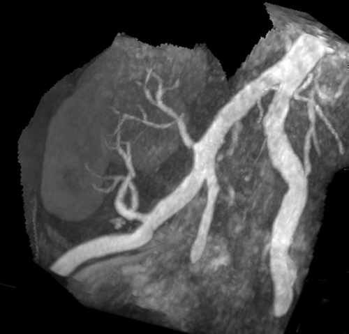
3D PC MR angiogram
|
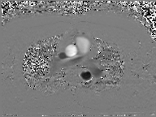
PC flow mapping at aortic root
|
|
The most commonly used applications today for phase-contrast imaging are: 1) in 2D mode to create a quick vascular scout image prior to performing a non-PC MRA technique; 2) in cine mode to quantify blood flows and velocities within the heart and great vessels; 3) in cine mode for qualitative or quantitative CSF flow measurements; 4) in 2D or 3D mode for intracranial MR venography; and 5) as a 3D MR angiogram for niche applications including MRA of the renal arteries and intracranial MRA in the presence of hemorrhage. The use of 3D-PC-MRA as a general angiographic method has greatly declined due to its long acquisition times and the availability of newer sequences (SSFP, FSE, and ASL) to perform the same function.
References
Bryant DJ, Payne JA, Firmin DN, et al. Measurement of flow with NMR imaging using a gradient pulse and phase difference technique. J Comput Assist Tomogr 1984;8:588-93.
Dyverfeldt P, Bissell M, Barker AJ, et al. 4D flow cardiovascular magnetic resonance consensus statement. J Cardiovasc Magn Reson 2015; 17:72.
Firmin DN, Nayler GL, Klipstein RH, et al. In vivo validation of MR velocity imaging. J Comput Assist Tomogr 1987;11:751-6.
Lotz J, Meier C, Leppert A, Galanski M. Cardiovascular flow measurement with phase-contrast MR imaging: basic facts and implementation. Radiographics 2002; 22:651-671.
Markl M, Frydrychowicz A, Kozerke S, et al. 4D Flow MRI. J Magn Reson Imaging 2012; 36:1015-1036.
Moran PR. A flow velocity zeugmatographic interlace for NMR imaging in humans. Magn Reson Imaging 1982; 1:197-203.
Moran PR, Moran RA, Karstaedt N. Verification and evaluation of internal flow and motion. True magnetic resonance imaging by the phase gradient modulation method. Radiology 1985;154:433-441.
Nayak KS, Nielsen J-F, Bernstein MA, et al. Cardiovascular magnetic resonance phase contrast imaging. J Cardiovasc Magne Reson 2015; 17:71.
Nayler GL, Firmin DN, Longmore DB. Blood flow imaging by cine magnetic resonance. J Comput Assist Tomogr 1986;10:715-22.
Stankovic Z, Allen BD, Garcia J, et al. 4D flow imaging with MRI. Cardiovasc Diagn Ther 2014; 4:173-192.
Wildermuth S, Debatin JF, Huisman TAGM, et al. 3D phase contrast EPI MR angiography of the carotid arteries. J Comput Assist Tomogr 1995; 19:871-878.
Related Questions
What are spin-phase effects?
What are spin-phase effects?