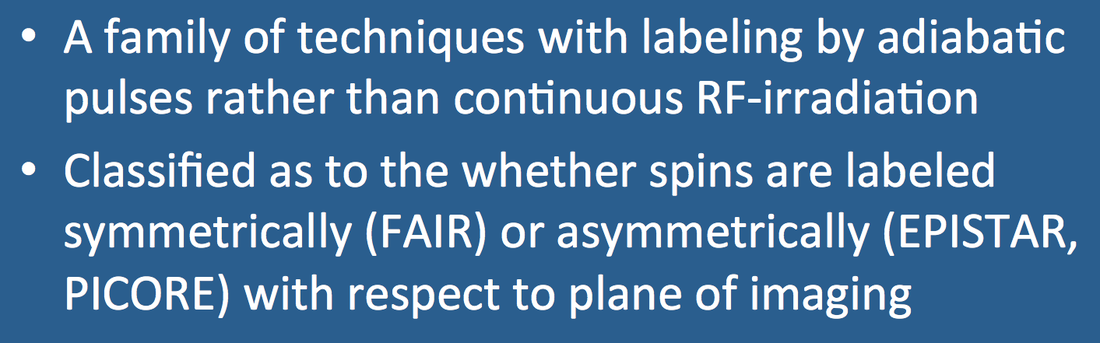
Pulsed Arterial Spin Labeling (PASL)
What is PASL and how does it differ from CASL?
|
|
Continuous Arterial Spin Labeling (CASL) techniques, described in the prior Q&A, are now primarily of historical and didactic interest, having fallen out of favor by the late 1990s. The main problems with CASL were its high tissue energy deposition and transmitter duty-cycle requirements accompanying the use of continuous RF-pulses. Thus came PASL, a family of ASL methods using pulsed (rather than continuous) RF-excitation. Three PASL variants are illustrated in the diagram below.
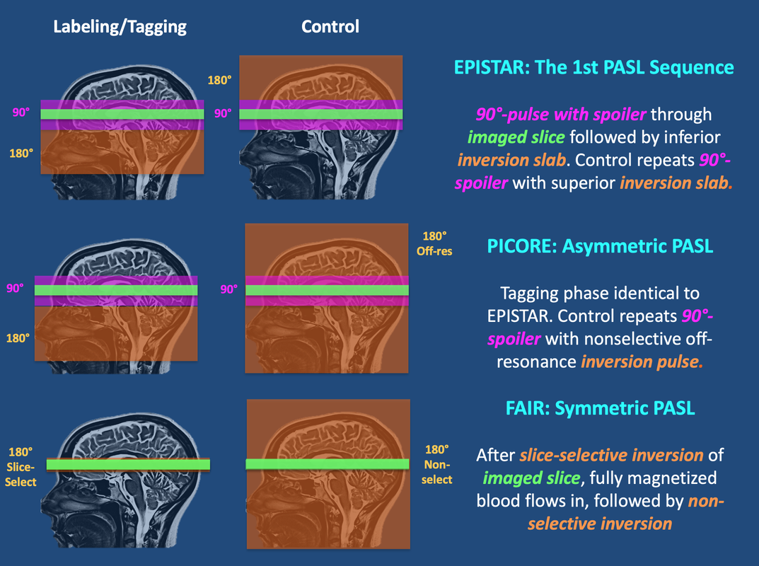
Pulsed Arterial Spin Labeling (PASL) methods
The first PASL method was developed by Edelman and colleagues in the mid-1990s and called EPISTAR (Echo-Planar Imaging-based Signal Targeting by Alternating Radiofrequency pulses). The tagging sequence of EPISTAR begins with a slice-selective 90°-pulse and spoiler gradient to initially saturate magnetization in the imaged slice. (The profile of this pulse is usually slightly wider than the imaged slice to minimize artifacts from RF-side lobes and ensure uniform saturation). A wide inversion slab is then applied proximally using a 180° adiabatic RF-pulse that inverts inflowing spins. The control sequence repeats the 90°-spoiler saturation of static tissue in the imaged slice, followed by a mirror-image distally placed thick inversion slab to create MT effects equivalent to those of the first slab. Subtraction of the tagged from control images produces a perfusion-weighted image where MT effects have been minimized.
Single-slice EPISTAR has subsequently evolved into several multi-slice variants (STAR, PULSAR, and QUASAR), some of which have been offered as commercial products by Philips. These are described briefly in the Advanced Discussion.
PICORE (Proximal Inversion with Control of Off-Resonance Effects) is another asymmetric multislice PASL technique with similarities to EPISTAR. In fact, the tagging sequence is identical to EPISTAR with imaging slice saturation and proximal inversion pulses. The control sequence differs in that the 180°-inversion pulse is applied at the same frequency offset relative to the imaging slice as the tagging sequence, but in the absence of a spatial gradient.
FAIR (Flow-sensitive Alternating Inversion Recovery) uses a slightly different pulsed labeling strategy that is symmetric with respect to the imaging slice. The tagged sequence begins with a spatially selective inversion pulse limited to a small region in and around the imaged slice. (The inversion pulse is usually slightly wider than the imaged slice to minimize artifacts from RF-side lobes and ensure uniform inversion). After this inversion pulse, "fresh" (fully magnetized) blood flows into the slice which serves as the "tag". This is followed by a non-slice-selective inversion pulse to the entire imaged volume as the "control". Versions of both PICORE and FAIR with Q2-TIPS bolus saturation (see Advanced Discussion) are currently offered as Siemens ASL products.
FAIR (Flow-sensitive Alternating Inversion Recovery) uses a slightly different pulsed labeling strategy that is symmetric with respect to the imaging slice. The tagged sequence begins with a spatially selective inversion pulse limited to a small region in and around the imaged slice. (The inversion pulse is usually slightly wider than the imaged slice to minimize artifacts from RF-side lobes and ensure uniform inversion). After this inversion pulse, "fresh" (fully magnetized) blood flows into the slice which serves as the "tag". This is followed by a non-slice-selective inversion pulse to the entire imaged volume as the "control". Versions of both PICORE and FAIR with Q2-TIPS bolus saturation (see Advanced Discussion) are currently offered as Siemens ASL products.
The choice between EPISTAR, PICORE, and FAIR methods should depend in part on the geometry of expected blood flow to the imaging volume. PICORE tags blood exclusively from one side and is thus a reasonable choice for axial brain perfusion imaging where all supply is coming from the neck. In cases where inflow may be from multiple or unknown directions (such as for sagittal brain perfusion imaging or in a complex organ such as the liver), FAIR may be a better choice since it tags both sides of the imaging volume. Conversely, FAIR may be more sensitive to contamination by unwanted venous inflow and is somewhat more difficult to implement in multi-slice mode due to inversion slice-profile imperfections. In some situations EPISTAR may be advantageous to the other methods having fewer eddy current artifacts due to its balanced slab-select gradients between control and tagged sequences.
References
Edelman RR, Chen Q. EPISTAR MRI: Mulitslice mapping of cerebral blood flow. Magn Reson Med 1998; 40:800-805. (Later iteration of EPISTAR with modifications and multi-slice acquisition)
Edelman RR, Siewert B, Darby DG, et al. Qualitative mapping of cerebral blood flow and functional localization with echo-planar MR imaging and signal targeting with alternating frequency. Radiology 1994; 192:513-520. (one of the early descriptions of EPISTAR)
Golay X, Hendrikse J, Lim TCC. Perfusion imaging using arterial spin labeling. Top Magn Reson Imaing 2004; 15:10-27. (Good review plus a description of ASL of variants and acronyms such as FAIRER, TILT, BASE, and others).
Golay X, Peterson ET, Hui F. Pulsed Star labeling of arterial regions (PULSAR): a robust regional perfusion technique for high field imaging. Magn Res Med 2005; 53:15-21.
Kim S-G. Quantification of relative cerebral blood flow change by Flow-sensitive Alternating Inversion Recovery (FAIR) technique: application to functional mapping. Magn Reson Med 1995; 34:293-301. (original description of FAIR technique)
Luh W-M, Wong EC, Bandettini PA, Hyde JS. QUIPSS II with thin-slice TI1 periodic saturation: a method for improving accuracy of quantitative perfusion imaging using pulsed arterial spin labeling. Magn Reson Med 1999; 41:1246-1254 (description of the Q2TIPS method).
Petersen ET, Lim T, Golay X. Model-free arterial spin labeling quantification: approach for perfusion MRI. Magn Reson Med 2006; 55:219-232. (QUASAR method)
Wong EC, Buxton RB, Frank LR. Implementation of quantitative perfusion imaging techniques for functional brain mapping using pulsed arterial spin labeling. NMR in Biomed 1997; 10:237-249. (description of the PASL/PICORE method)
Related Questions
Can you briefly explain the difference between the various ASL methods? Which is the best?
What is pCASL and how does it differ from CASL and PASL?
Can you briefly explain the difference between the various ASL methods? Which is the best?
What is pCASL and how does it differ from CASL and PASL?